Vitreo Retinal Surgery in Mumbai(Vitreo Retinal superspeciality department)at the Wavikar Eye institute is capable of handling complicated Retina disorders.
With the emerging need to attend to retinal problems effectively,we also believe in prophylactic treatment for various early stages retinal problems.Early detection and early intervention is also helpful to prolong the rapid deterioration of the vision.

Diabetes: – Diabetes is a disease characterized by increased sugar level in blood either because of inadequate secretion of insulin or improper processing of if by the body.
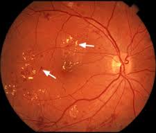
Diabetic retinopathy is one of the vascular (Blood-vessel related) complications related to diabetes.It is due to damage of small vessels and so is called a “microvascular complication”.Kidney disease and nerve damage due to diabetes are also microvascular complications. Large blood vessel damage (also called macro vascular complications) includes heart disease and stroke.
Diabetes retinopathy is the leading cause irreversible blindness in industrialized nationsif retinopathy is not found early or is not treated,it can lead to blindness.
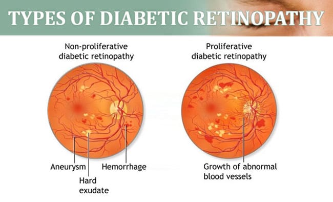
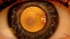
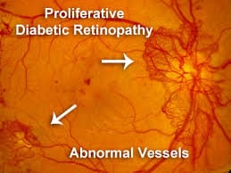
HERE, IT IS IMPORTANT TO ADDRESS THE RISKS FACTORS THAT CAN WORSEN THE OCCLUDED VESSELS. SMOKING CESSATION, HYPERTENSION CONTROL, CHOLESTEROL MANAGEMENT AND GLUCOSE CONTROL MUST TAKE PLACE IN ORDER TO STOP THE PROGRESSION OF NEW VESSELS FROM FORMING.
ANNUAL EYE CHECK UP IS MANDATORY FOR ALL DIABETIC PATIENTS:-
ONCE THE SYMPTOMS ARISE IMMEDIATE CHECK UP & MORE FREQUENT FOLLOW UPS ARE NECESSARY. SYMPTOMS OF DIABETIC RETINOPATHY ARE BLACK SPOTS IN YOUR VISION, FLASHES OF LIGHT, HOLES IN YOUR VISION & BLURRED VISION.
EXAMINATION OF THE EYE AFTER DILATATION OF PUPIL BY OPHTHALMOSCOPE.
FFA (FUNDS FLUORESCEIN ANGIOGRAPHY ):-METHOD OF EVALUATING THE PATENCY OF RETINAL AND CHOROIDAL BLOOD VESSELS, FLUORESCEIN DYE IS INJECTED INTO A VEIN THEN SUBSEQUENTLY PHOTOGRAPHS OF THE EYE ARE TAKEN I.E. PHOTOGRAPHS OF FUNDUS AS THE DYE CIRCULATES ARE TAKEN.
ULTRASONOGRAPHY (B ASCAN) OF THE EYE.TRANSMISSION OF HIGH FREQUENCY SOUND WAVES INTO THE EYES, WHICH ARE REFLECTED THE OCULAR TISSUES AND DISPLAYED ON SCREEN SO THAT INTERNAL STRUCTURES CAN BE VISUALIZED IN EYE WITH OPAQUE MEDIA.ULTRASONOGRAPHY INDICATES THE CHANGES THAT ARE TAKING PLACE IN THE VITREOUS HEMORRHAGE, DENSE CATARACT,AND CORNEAL OPACITY.B SCAN HELPS US TO EVALUATE SUCH EYES AND PLAN FOR SURGERIES.
ICG: – ANOTHER TYPE OF ANGIOGRAPHY OF THE VESSELS IN THE I C G (INDOCYANINE GREEN IS A DYE THAT LIGHTS UP WHEN EXPOSED TO INFRARED LIGHT.
INFRARED LIGHT IS USED TO TAKE PICTURES OF THE BACK OF THE EYE VISUALIZING RETINAL BLOOD VESSELS AND CHOROIDAL BLOOD VESSELS.
OCT: – OCT USES LIGHT WAVES SLICING THROUGH TISSUE LAYERS IN THE BACK OF THE EYE THEY PRODUCE A BACK SCATTERING THAT CONVERTS INTO HIGH RESOLUTION CROSS SECTIONAL IMAGES OF THE RETINA, MACULA AND OPTIC NERVE, WITH THIS NEW MEDICAL IMAGING TECHNOLOGY NERVE, WITH THIS NEW MEDICAL IMAGING TECHNOLOGY CLINICIANS HAVE ANOTHER PAINLESS, PRECISE DIAGNOSTIC TOOL.
LASER STANDS FOR LIGHT AMPLIFICATION BY STIMULATED EMISSION OF RADIATION.
LASER BEAM IS ENERGY THAT COMES FROM SPECIAL LIGHT SOURCE;IT IS FOCUSED ON TO THE DISEASED AREA IN THE RETINA. THE HEAT ENERGY SEALS AND DESTROYS THE ABNORMAL BLOOD VESSELS.THE AIM OF THIS TREATMENT IS TO PROTECT THE PATIENT’S MOST IMPORTANT CENTRAL VISION,SACRIFICING IF NECESSARY SOME OF THE LESS IMPORTANT SIDE VISION.
THE LASER TREATMENT STABILIZES THE PROGRESSION OF DIABETIC RETINOPATHY, PERIODIC FOLLOW UP IS IMPORTANT TO CHECK ANY FURTHER PROGRESSION IS THERE.
ONE HAS TO REMEMBER THAT VISION LOST DUE TO DIABETES IS VERY DIFFICULT TO RESTORE.
THE DOCTOR MAY ASK FOR A FUNDUS FLUROSCEIN ANGIOGRAPHY TO BE DONE BEFORE DECIDING ABOUT THE LASER BECAUSE AS IT ENABLES THE DOCTOR TO EVALUATE THE LEAKAGE IN THE RETINA.
LASER TREATMENT IS PAINLESS PROCEDURE. IN MOST CASES ARE AN OPD PROCEDURE AND NOT A SURGICAL PROCEDURE.
EXTENT OF DIABETIC RETINOPATHY PLAYS AN IMPORTANT ROLE IN DECIDING THE NUMBER OF SITTINGS REQUIRED.
YOU COULD HAVE BLURRED VISION, MILD WATERING, REDNESS AFTER LASER TREATMENT, THEY ARE USUALLY TEMPORARY.
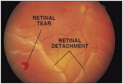
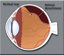
VITRECTOMY IS ANOTHER SURGERY COMMONLY NEEDED FOR DIABETIC PATIENTS WHO SUFFER A VITREOUS HEMORRHAGE (BLEEDING IN THE GEL-LIKE SUBSTANCE THE FILLS THE CENTER OF THE EYE).DURING A VITREO RETINAL SURGERY IN MUMBAI, THE RETINAL SURGEON CAREFULLY REMOVES BLOOD AND VITREOUS FROM THE EYE AND REPLACES IT WITH CLEAR SALT SOLUTION (SALINE). PATIENTS WITH DIABETES ARE AT GREATER RISK OF DEVELOPING RETINAL FEARS.AND DETACHMENT,TEARS IS OFTEN SEALED WITH LASER SURGERY. RETINAL DETACHMENT REQUIRES SURGICAL TREATMENT TO REATTACH THE RETINA TO THE BACK OF THE EYE. THE PROGNOSIS FOR VISUAL RECOVERY IS DEPENDENT ON THE SEVERITY OF THE DETACHMENT.
EVEN THOUGH RETINOPATHY CANNOT BE ELIMINATED COMPLETELY. STILL THE SEVERITY OF THE DAMAGE CAN BE MINIMIZED, IF DIABETES IS WELL WITHIN CONTROL AND IF THE TREATMENT HAS BEEN STARTED EARLY.
IN MORE THAN 60% OF CASES, THE VISION CAN BE SAVED IF THE TREATMENT WITH LASER HAS BEEN GIVEN IN TIME & ELIMINATE THE NEED FOR MORE COMPLICATED SURGICAL PROCEDURE.
WAVIKAR EYE INSTITUTE PROVIDES BEST VITRO RETINAL SURGERY IN MUMBAI.